How Does Silicone Gel Work on Scars? Mechanisms and Clinical Evidence
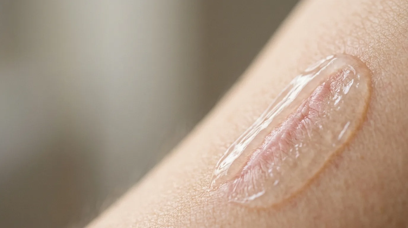
Topical silicone works by providing a breathable, semi-occlusive barrier that reduces transepidermal water loss and restores stratum corneum hydration. This normalized microenvironment signals dermal fibroblasts to downregulate excessive collagen production, thereby helping to soften, flatten, and smooth the overall appearance of healing scar tissue.
Following cutaneous injury, surgical intervention, or trauma, the human integument initiates an intricate repair cascade to re-establish tissue continuity. When deep dermal architecture is breached, the final phase—maturation and remodeling—often results in noticeable textural irregularities, heightened erythema, or raised contours. Understanding how does silicone gel work on scars requires examining the biophysical environment of healing tissue, particularly the interplay between stratum corneum barrier integrity, water retention, and dermal cellular communication.
Topical silicone formulations have been evaluated extensively across cutaneous research for decades. Unlike pharmacological agents that penetrate systemically to alter cellular pathways directly, topical silicone behaves primarily as a non-reactive physical barrier. By modulating the microclimate of the healing epidermis, it creates physiological conditions that guide endogenous remodeling toward a softer, flatter, and more compliant tissue state.
What Biological Mechanisms Drive Silicone's Impact on Scars?
During normal epidermal maturation, intact stratum corneum cells and intercellular lipid matrices regulate transepidermal water loss (TEWL). When skin sustains full-thickness injury, re-epithelialization produces an immature, fragile epidermal layer that lacks complete barrier competence. Consequently, TEWL through newly formed skin can remain substantially elevated for months after initial wound closure.
Elevated water evaporation signals persistent barrier dysfunction to the underlying viable epidermis. Keratinocytes respond to chronic dehydration by releasing inflammatory mediators and profibrotic cytokines. These signaling cascades prompt underlying dermal fibroblasts to sustain elevated cellular activity, leading to dense, disorganized extracellular matrix deposition characteristic of hypertrophic scars and persistent erythema.
Topical silicone agents function through semi-occlusive barrier restoration, which moderates excessive fibroblast signaling and excessive collagen deposition in healing tissues 29601619. When applied across the immature scar surface, silicone polymers form a continuous, flexible layer that is permeable to gases such as oxygen and carbon dioxide, yet semi-impermeable to liquid water. This physical behavior mimics the moisture-retentive capacity of healthy stratum corneum, sharply curbing excessive TEWL without causing epidermal maceration.
Restoring stratum corneum hydration normalizes the osmotic state within epidermal cells. Once local dehydration stress subsides, keratinocytes downregulate the transmission of growth factors that otherwise stimulate excessive extracellular matrix synthesis. As dermal fibroblast hyperactivity recedes, the balance between matrix accumulation and enzymatic remodeling stabilizes, allowing disorganized structural fibers to align more harmoniously over time.
Exactly How Does Silicone Gel Work on Scars at the Microscopic Level?
To appreciate how does silicone gel work on scars during active tissue remodeling, one must trace the cellular events connecting surface hydration to deep dermal organization. The primary site of action is not deep within the subcutaneous tissue; rather, the biological cascade initiates entirely at the outermost interface.
Under unoccluded conditions, an immature scar continues to lose water at rates up to four times higher than unwounded skin. This continuous fluid flux creates a persistent state of cellular dehydration in the stratum granulosum and stratum spinosum. In response to this hyperosmotic stress, epidermal cells synthesize and release signaling peptides, including interleukin-1 alpha and transforming growth factor-beta. These molecules diffuse across the dermo-epidermal junction into the papillary and reticular dermis.
In the dermis, resident fibroblasts intercept these epidermal distress signals. Fibroblasts interpret sustained cytokine flux as an ongoing regenerative emergency, causing them to deposit dense bundles of extracellular matrix components and form irregular aggregates. This continuous deposition creates the palpably rigid, elevated profile recognized in hypertrophic scars. Furthermore, persistent subclinical inflammation sustains local microvascular proliferation, presenting clinically as prolonged erythema.
Applying a topical silicone gel directly interrupts this cycle. The silicone polymer cross-links upon ambient drying to yield an ultra-thin, resilient film. By capturing escaping water vapor, the polymer increases hydration within the stratum corneum, effectively restoring turgor and normal biochemical homeostasis within the epidermis. Relieved of osmotic strain, keratinocytes cease the sustained production of profibrotic cytokines. In the absence of sustained activation signals, fibroblasts scale back the excessive synthesis of matrix components, and local capillary hyper-reactivity subsides. Over progressive weeks of consistent coverage, this cellular normalization supports a visibly flatter, less erythematous, and more pliable surface.
How Do Silicone Gels and Silicone Sheets Differ in Application?
Topical silicone is traditionally provided in two physical presentations: elastomeric silicone sheet dressings and self-drying fluid silicone gels. Both share identical foundational mechanisms—namely, semi-occlusive hydration and TEWL reduction—yet their practical handling characteristics suit different clinical contexts.
A randomized controlled trial on postoperative scar management confirmed that silicone sheeting significantly reduced scar thickness and improved clinical scores compared to unoccluded controls 27538109. Silicone sheets are adhesive, rubber-like pads designed to be applied directly onto planar skin areas, such as the sternum, abdomen, or extremities. Beyond providing barrier hydration, sheets offer modest mechanical stabilization by splinting wound edges against ambient shearing forces and mechanical tension. However, sheets require diligent daily cleaning, may loosen under perspiration, and can be visually conspicuous on visible body sites.
Conversely, self-drying fluid silicone gels were developed to overcome anatomical limitations associated with solid sheeting. Fluid gels consist of long-chain silicone polymers blended with volatile silicone solvents. Upon spreading a micro-thin layer across the skin, the volatile carrier evaporates within minutes, leaving behind an invisible, gas-permeable film that conforms precisely to irregular contours.
Fluid gels offer clear advantages when managing scars across mobile anatomical regions, including joints, facial structures, and curved periorbital contours. Unlike solid dressings, fluid films do not pull on delicate tissues, lift at the margins during facial expression, or cause perimeter maceration. Both formats demonstrate documented utility in supporting scar maturation and pliability when used consistently, allowing healthcare professionals to select the presentation best aligned with anatomical location and daily routine.
What Does Clinical Evidence Reveal About Post-Intervention Scar Maturation?
Rigorous clinical research has consistently validated the physiological utility of topical silicone in post-wound skin recovery. Modern controlled trials demonstrate that early intervention following complete re-epithelialization substantially improves visual and tactile parameters of newly formed tissue.
In a randomized, controlled, double-blind trial evaluating postoperative eyelid incisions, topical silicone gel demonstrated statistically significant aesthetic improvements compared to placebo 39946309. Periocular and eyelid scars represent a uniquely sensitive anatomical challenge due to extreme tissue thinness, rapid blinking dynamics, and high visibility. The study observed that protocolized, twice-daily application of silicone gel led to measurable enhancements in overall scar score, vascularity grading, and scar height compared to vehicle control, confirming that fluid polymers perform reliably even in complex, delicate facial zones.
Similarly, comparative evaluations have analyzed silicone performance following deliberate procedural skin injury. A split-face comparative study demonstrated that silicone-based fluid gels provide effective barrier support and wound recovery following fractional ablative laser trauma 41195573. In this clinical setting, high-energy laser columns disrupt the stratum corneum across microscopic zones, requiring robust, non-irritating barrier restoration to mitigate prolonged post-procedure erythema and dyschromia. Patients treated with fluid silicone gels experienced favorable barrier recovery without the occlusive folliculitis or comedogenic drawbacks occasionally observed with heavy petrolatum-based ointments.
Collectively, these controlled investigations demonstrate that protocolized topical silicone supports the natural phases of tissue remodeling. Whether applied following sharp surgical excision or energy-based thermal resurfacing, continuous semi-occlusive management preserves hydration, mitigates excessive vascular response, and fosters optimal aesthetic integration with surrounding healthy skin.
Practical Protocol: When and How to Apply Topical Silicone Formulations
Achieving the physiological benefits of topical silicone requires meticulous adherence to application timing and technique. Because silicone acts mechanically rather than systemically, therapeutic outcomes depend on continuous contact during the vulnerable remodeling window.
Application must begin only after the wound has achieved complete re-epithelialization. This means that surgical incisions must be fully closed, initial scabbing must have sloughed off naturally, and any sutures or surgical adhesives must be fully removed by a physician. Applying silicone over open wounds, unhealed abrasions, or broken skin is inappropriate and may impede natural cellular migration.
Prior to application, the skin should be gently cleansed with a mild, non-drying cleanser and patted completely dry. A very small quantity of silicone gel should be smoothed across the tissue in a thin, even layer. Over-application does not enhance performance; excessive gel simply prolongs drying time and increases the likelihood of transfer onto clothing. A properly applied thin layer self-dries within three to five minutes, forming a dry, non-greasy, flexible film. Once fully dry, appropriate high-protection broad-spectrum sun care may be layered on top to safeguard healing skin from UV-induced hyperpigmentation.
Consistency represents the most decisive factor in scar management protocols. Clinical observations indicate that topical silicone should remain in contact with the skin for virtually the entire day—ideally between 12 and 24 hours daily. Because connective tissue remodeling is an intrinsically slow physiological process, visible improvements in scar softness, redness, and height typically emerge between 8 to 12 weeks of uninterrupted application. For mature or unusually resistant scars, healthcare providers often advise maintaining the regimen for up to six months to achieve optimal structural stabilization.
Key takeaways
- Topical silicone establishes a breathable, semi-occlusive barrier that reduces transepidermal water loss and normalizes stratum corneum hydration.
- Restored epidermal hydration downregulates profibrotic cytokine signaling, preventing dermal fibroblasts from depositing excessive extracellular matrix.
- Silicone gel conforms readily to dynamic facial and joint contours where solid silicone sheets are difficult to maintain.
- Controlled clinical trials confirm that topical silicone significantly reduces postoperative scar height, erythema, and overall scar scores.
- Application must commence only after complete re-epithelialization and must continue consistently for 8 to 12 weeks for noticeable refinement.
Frequently asked questions
- When is the optimal time to initiate topical silicone application after skin re-epithelialization?
- Topical silicone should be initiated immediately after the skin barrier has completely closed, scabs have shed naturally, and all surgical sutures have been removed. It must never be applied to open, exuding, or incompletely healed wounds.
- How do silicone gels compare with silicone sheets for mobile or facial areas?
- Silicone gels dry to form an invisible, flexible film that adheres easily to mobile facial contours, eyelids, and joint areas without peeling. Silicone sheets are better suited for flat, non-mobile areas such as the torso or limbs.
- How many weeks of continuous application are typically required to notice visible textural changes in scars?
- Because dermal remodeling occurs slowly, visible changes in scar pliability, height, and erythema generally require 8 to 12 weeks of continuous, daily application. Prolonged use up to six months may be advised by a physician for stubborn tissue.
- Does topical silicone assist with both new surgical scars and older, mature tissue?
- While topical silicone exhibits its greatest clinical benefit during active remodeling in newer scars, it can also help soften and improve the overall pliability and surface appearance of mature, older scars, though responses typically require longer application periods.
References
- Efficacy of Silicone Gel Versus Placebo for Postsurgical Scars of the Eyelids: A Randomized, Controlled, Double-Blinded Study — Ophthalmic plastic and reconstructive surgery · 2025
- The Efficacy of a Silicone Sheet in Postoperative Scar Management — Advances in skin & wound care · 2016
- Topical Agents for Scar Management: Are They Effective? — Journal of drugs in dermatology : JDD · 2018
- Silicone-based fluid gel versus white paraffin ointment in the treatment of post-fractional ablative CO(2) laser wound: an intra-individual split-face comparative study — The Journal of dermatological treatment · 2025

This content is for information only and does not replace a medical consultation. Consult your doctor about your own situation.